
ROBERT HASTEROK
University of Silesia,
Faculty of Biology and Environmental Protection, Department of Plant Anatomy and Cytology;
ul. Jagiellońska 28, 40-032 Katowice,
e-mail: robrert.hasterok@us.edu.pl
Description popularizing the research project
First we can observe a tiny white root which immediately bends downward following the direction of gravity. It is growing fast. It is quickly followed by a green shoot with seed leaves. Within a few days a batch of sprouts for salad is ready. Within the days in the tasty sprout cells replicate. They grow and divide, grow and divide. Each one splits into two others. And all that happens at a really breathtaking speed. “Pity that you can't see itł" you would like to exclaim. It must be truly fascinating although it does not resemble a football match, but rather synchronized swimming.
Since the very beginning cells replicate through division. They split in halves. Each of the daughter cells is a clone of its mother, the same in each aspect as its twin sister. Identical twin sister we could say. Identical both externally, on the surface of cell walls, and internally, in the nucleus which is both heart and brain of the cell. It is where the chromosomes, gene carriers, get their characteristic rod-shaped form with a constriction in the middle. It is where the rods find their pairs in a way which cannot be accidental and form a double line. After a while the Prussian drill turns into a colorful march of a band of majorettes or a group of beauties involved in synchronized swimming in cytoplasm. The characteristic colors of certain fragments of chromosomes and almost pitch black background give the picture even more charm. Thanks to the precise choreography we have healthy food for breakfast, fields of corn around, mosquitoes, swallows and our own limbs.
Abstract
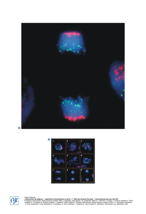
The aim of this project was to visualise the course of mitotic division in nuclei of root-tip meristematic cells in of rye (Secale cereale) using fluorescent in situ hybridisation (FISH), a modern molecular cytogenetics technique in which genes or non-coding DNA sequences can be detected with a microscope. The sequences of interest are visualised by hybridisation to complementary, fluorescently labelled DNA particles called probes. In this experiment probes for centromeres (red fluorescence) and telomeres (green fluorescence) highlight the elegance and dynamics of the division of chromosomal DNA (blue fluorescence). In interphase (Fig. 1) chromosomes replicate into two chromatids but are not yet visible individually. Prophase (Fig. 2) is the very first moment when chromosomes contract and appear as threads. In early metaphase (Fig. 3) chromosomes migrate to the equator of the spindle and at late metaphase (Fig. 4) orientate themselves ready for sister chromatid segregation. Treatment with colchicine (Fig. 5) at this point destroys the spindle and hypercondenses the chromatin. It is very useful for counting and identifying individual chromosomes. In early anaphase (Fig. 6) chromatids segregate to opposite poles, where they start to decondense (Fig. 7). During late anaphase (Fig. 8) individual chromosomes can no longer be distinguished. Finally, in telophase (Fig. 9) two identical daughter nuclei are produced from one parental cell.