2006
2006
 2008
2008
 2010
2010
 2012
2012
 2014
2014


 |
"Proteins in gel – or how to set… a laboratory table"
‘Proteomics of barley roots – optimization of protein extraction method from barley seedlings roots, for the use in 2D electrophoresis’
Katarzyna Bzdęga1 – author of photograph ,
2University of Silesia in Katowice, Faculty of Biology and Environmental Protection,
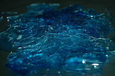
‘Moon landscape of gels’ – residues of gel scanning |

|
Description popularizing the research project
‘… what is essential is invisible to the eye’ once claimed Antoine de Saint Exupery. Surprisingly the quote about life may refer to proteomics as well. There are proteins which can be observed without the state-of-art equipment, complex procedures and specialist knowledge e.g. haemoglobin. Everybody recognizes its ruby red colour. The same is with luciferin. The glow emitted by fireflies and rotten wood betrays its presence. It is hard to miss when it starts shining on the bottom of a forest. But there are only a few such spectacular proteins. The vast innumerable majority of these important biomolecules remains hidden from the view and the symptoms of their presence are not conspicuous. Yet because of various functions they play, e.g. in shaping qualities and characteristics of organisms, it is crucial to be able to detect and describe them. Especially the ones that are created in cells of the organisms that, for various reasons, are important for health or life, or play a significant role in human economy. The latter ones are proteins of such plants as barley, fodder, ornamental plants, or domesticated animals. Yet many of the proteins often avoid detection – they mimic other protein compounds, disappear in a crowd of similar particles, link in pairs, quartets or even bigger agglomerates of particles, pretending to be bigger molecules. All of that makes their detection a challenge for researchers. Yet nowadays use of an appropriate method allows obtaining an image of all the proteins present in given tissue or body organ at a certain development stage. They are worth looking for – description of the imaged proteins and learning their qualities opens a new door for science and often provides biotechnology and economy with new tools. It is necessary to look into “the invisible” to understand the visible signs of proteins – mysterious biomolecules so important in our everyday life.
Abstract
Proteomics allows for global analysis of proteins accumulated in specific tissue during specific developmental time. The techniques that are used allow studying the changes in the level of protein accumulation and discovering functional links between different proteins. Proteomics gives also the possibility to study both structural proteins and enzymes, which directly reflects a specific phenotypic effect. Such a correlation is not always true for the studies of gene expression. The main goal of the project was the development of efficient and repetitive method of protein extraction, which will give high quality extracts adjusted for the two-dimensional (2D) electrophoresis. The material of the study was roots from 6-day-old barley seedlings growing in aeroponic conditions, which were collected and ground in liquid nitro-gen. Proteins were extracted from 100 mg of material using three methods: • extraction with phenol, • extraction by precipitation in trichloroacetic acid (TCA), • extraction with the use of TriPure reagent. After 2D electrophoresis gels were scanned and analyzed taking into consideration number of spots and quality of electrophoresis – the appearance of vertical or horizontal smear and gel background, the evidence of impurities in the protein extracts. The best results were obtained using extraction with TCA, which were further optimized by using 250 mg of tissue and the addition of hydroxyethyl disulfide. This reagent prevents the unspecific oxidation of protein thiol groups which prevents formation of horizontal smears – the result of imprecise separation of proteins according to their isoelectric points. After gel image analysis 769 good quality spots were found which allowed to start the studies of barley root proteome.
Komitet Organizacyjny


Sponsorzy


Patronat honorowy
Leszek Jodliński
Dyrektor Muzeum Śląskiego w Katowicach
Zygmunt Łukaszczyk
Wojewoda Śląski
Jan Malicki
Biblioteka Śląska
Piotr Uszok
Prezydent Katowic
Adam Matusiewicz
Marszałek Województwa Śląskiego
Patronat medialny